[Advanced Published online The Keio Journal of Medicine, by J-STAGE]
<Title:> Visualization of Lymphatic Vessels Using Photoacoustic Imaging
<Author(s):> Hiroki Kajita, Yushi Suzuki, Hisashi Sakuma, Nobuaki Imanishi, Tetsuya Tsuji, Masahiro Jinzaki, Sadakazu Aiso, Kazuo Kishi
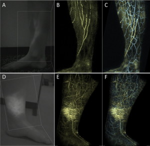
<Abstract:> Lymphedema occurs when interstitial fluid and fibroadipose tissues accumulate abnormally because of decreased drainage of lymphatic fluid as a result of injury, infection, or congenital abnormalities of the lymphatic system drainage pathway. An accurate anatomical map of the lymphatic vasculature is needed not only for understanding the pathophysiology of lymphedema but also for surgical planning. However, because of their limited spatial resolution, no imaging modalities are currently able to noninvasively provide a clear visualization of the lymphatic vessels. Photoacoustic imaging is an emerging medical imaging technique that provides unique scalability of optical resolution and acoustic depth of penetration. Moreover, light-absorbing biomolecules, including oxy- and deoxyhemoglobin, lipids, water, and melanin, can be imaged. Using exogenous contrast agents that are taken up by lymphatic vessels, e.g., indocyanine green, photoacoustic lymphangiography, which has a higher spatial resolution than previous imaging modalities, is possible. Using a new prototype of a photoacoustic imaging system with a wide field of view developed by a Japanese research group, high-resolution three-dimensional structural information of the vasculatures was successfully obtained over a large area in both healthy and lymphedematous extremities. Anatomical information on the lymphatic vessels and adjacent veins provided by photoacoustic lymphangiography is helpful for the management of lymphedema. In particular, such knowledge will facilitate the planning of microsurgical lymphaticovenular anastomoses to bypass the excess fluid component by joining with the circulatory system peripherally. Although challenges remain to establish its implementation in clinical practice, photoacoustic lymphangiography may contribute to improved treatments for lymphedema patients in the near future.
<Keywords:> photoacoustic techniques, lymphatic vessels, lymphedema, indocyanine green
<URL:> https://www.jstage.jst.go.jp/article/kjm/advpub/0/advpub_2020-0010-OA/_html
<Title:> Visualization of Lymphatic Vessels Using Photoacoustic Imaging
<Author(s):> Hiroki Kajita, Yushi Suzuki, Hisashi Sakuma, Nobuaki Imanishi, Tetsuya Tsuji, Masahiro Jinzaki, Sadakazu Aiso, Kazuo Kishi
<Abstract:> Lymphedema occurs when interstitial fluid and fibroadipose tissues accumulate abnormally because of decreased drainage of lymphatic fluid as a result of injury, infection, or congenital abnormalities of the lymphatic system drainage pathway. An accurate anatomical map of the lymphatic vasculature is needed not only for understanding the pathophysiology of lymphedema but also for surgical planning. However, because of their limited spatial resolution, no imaging modalities are currently able to noninvasively provide a clear visualization of the lymphatic vessels. Photoacoustic imaging is an emerging medical imaging technique that provides unique scalability of optical resolution and acoustic depth of penetration. Moreover, light-absorbing biomolecules, including oxy- and deoxyhemoglobin, lipids, water, and melanin, can be imaged. Using exogenous contrast agents that are taken up by lymphatic vessels, e.g., indocyanine green, photoacoustic lymphangiography, which has a higher spatial resolution than previous imaging modalities, is possible. Using a new prototype of a photoacoustic imaging system with a wide field of view developed by a Japanese research group, high-resolution three-dimensional structural information of the vasculatures was successfully obtained over a large area in both healthy and lymphedematous extremities. Anatomical information on the lymphatic vessels and adjacent veins provided by photoacoustic lymphangiography is helpful for the management of lymphedema. In particular, such knowledge will facilitate the planning of microsurgical lymphaticovenular anastomoses to bypass the excess fluid component by joining with the circulatory system peripherally. Although challenges remain to establish its implementation in clinical practice, photoacoustic lymphangiography may contribute to improved treatments for lymphedema patients in the near future.
<Keywords:> photoacoustic techniques, lymphatic vessels, lymphedema, indocyanine green
<URL:> https://www.jstage.jst.go.jp/article/kjm/advpub/0/advpub_2020-0010-OA/_html

![Acceptability and Preliminary Effects of Online Evidence-based Practice Education Program for Undergraduate Nursing Students: A Pre- and Post-intervention Study [Published online in advanced , by J-STAGE]](http://kjm.pupu.jp/blog/wp-content/uploads/2025/09/2024-0013-OA-100x100.jpg)
![Neurofibromatosis 1 (von Recklinghausen Disease) [Published online in advanced , by J-STAGE]](http://kjm.pupu.jp/blog/wp-content/uploads/2023/08/2023-0013-IR-100x100.jpg)
![Community Pharmacists’ Perceptions and Needs Regarding Oral Healthcare Advice in Japan [Published online in advanced , by J-STAGE]](http://kjm.pupu.jp/blog/wp-content/uploads/2025/05/2024-0022-OA-100x100.jpg)
![Potential New Tumors Associated with Hereditary Breast and Ovarian Cancer (HBOC) [Published online Keio J Med, 74, 158-161, by J-STAGE]](http://kjm.pupu.jp/blog/wp-content/uploads/2025/09/2024-0023-RE-100x100.jpg)
![Hypertrophic Cardiomyopathy: Diverse Pathophysiology Revealed by Genetic Research, Toward Future Therapy [Published online Keio J Med, 69, 77-87, by J-STAGE]](http://kjm.pupu.jp/blog/wp-content/uploads/2020/12/2019-0012-OA-100x100.jpg)
![Pachyonychia Congenita: Clinical Features and Future Treatments [Published online Keio J Med, 74, 52-60, by J-STAGE]](http://kjm.pupu.jp/blog/wp-content/uploads/2025/03/2023-0012-IR-100x100.jpg)
![A Case of Severe Rectal Hemorrhage Possibly Caused by Radiation Recall after Administration of Gemcitabine [Published online Keio J Med, 65, 16-20, by J-STAGE]](http://kjm.pupu.jp/blog/wp-content/uploads/2016/03/2014-0015-CR-100x100.jpg)
![Network Approaches to Uncover Pathogenesis and Therapeutic Targets of Inflammatory Bowel Diseases [Published online in advanced , by J-STAGE]](http://kjm.pupu.jp/blog/wp-content/uploads/2023/03/2022-0015-IR-100x100.jpg)
![Development of Cardiac Regenerative Medicine Using Human iPS Cell-derived Cardiomyocytes [Published online in advanced , by J-STAGE]](http://kjm.pupu.jp/blog/wp-content/uploads/2020/08/2020-0009-IR-100x100.jpg)